Radiology
You have been advised by your doctor to have an MRI arthrogram. This leaflet will explain what the examination involves. Please read this leaflet carefully to ensure you successfully prepare for the examination.
This test is in great demand and we would be grateful if you could ensure that you arrive for your appointment in plenty of time. If for any reason you are unable to keep your appointment, please let us know as soon as possible.
What is an Arthrogram and Magnetic Resonance Imaging Scan (MRI)
This diagnostic test is an MRI scan, but in order to show up your joint properly, a special dye needs to be inserted directly into your joint using fluoroscopy (X rays).
Fluoroscopy
An arthrogram is a special X-ray procedure to look at the joints within the body such as the shoulders, wrists, hips or knees. The area to be examined will be cleaned and covered with a sterile drape. Local anaesthetic will be used to numb the area, which may sting a little at first. A very fine needle will be used to inject some contrast medium, which shows up on X rays, into the joint space. X rays are used during the procedure for guidance. This part of the examination will take approximately 15 minutes.
MRI is the next part of the test which uses a powerful magnet and a computer to produce detailed images of any part of the body. The MRI scanner does not use X-rays. This part of the examination will be performed by a radiographer and will take approximately 30 minutes.
What does the machine look like?
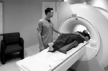
The equipment may look intimidating but there is no need to be nervous. The MRI scanner looks like a large elongated polo mint, which is well lit and open at both ends. The scanner remains open and you are never totally enclosed. Some scans require you to be placed head first in the scanner and others feet first.
What happens during the scan?
Throughout the procedure you will be asked to lie on a firm couch which moves in and out of the scanner, all you need to do is relax and stay still. You will be offered some earplugs to help reduce the noise of the scanner (a loud knocking noise).
Cautions
It is vital that you remove metallic objects before an MRI exam, including watches, jewellery, and items of clothing that have metallic threads, underwires or fasteners. It would be best to leave watches, jewellery or anything made from metal at home if possible.
You will also need to remove any bank cards and electrical items from your person such as mobile phones as the scanner will interfere with them and stop them from working.
You may prefer to arrive for your scan in clothes which do not contain metal. It will not be a problem if you need to get changed, we provide scrubs to change into if required.
Please complete the enclosed safety questionnaire and bring it with you to your appointment.
It is recommended that you do not drive for the remainder of the day so you will require someone to drive you to and from your appointment.
Medication
Please continue with your medication as normal. Please bring any sprays or inhalers that you are taking with you to your appointment.
What do I need to tell you about
For all patients between the ages of 12 and 55 who have ovaries, the x-ray department has a legal responsibility to ensure that this examination is performed within TEN DAYS of the first day of your menstrual period. Please contact the x-ray department if you are pregnant or if this appointment is beyond the TEN DAY requirement, and another appointment will be arranged for you.
Radiation dose and risk
Xray uses ionising radiation which can cause cell damage that may, after many years or decades, turn cancerous. The risk of this happening is very small compared to the lifetime risk of developing cancer which is 1 in 2. We are also all exposed to background radiation every day.
The risk of long-term effects is considered when the healthcare team decides whenever someone needs an X ray examination and radiation doses are kept as low as possible. For this examination radiation dose levels are typically equivalent to 6 to 12 months of background radiation. The associated risk is 1 in 10 000 – very low.
Contrast
The contrast agents are considered to be safe. As with all drugs and medication there is a slight risk of allergic reaction This may vary from a rash, to, very rarely, a more severe reaction.
Severe allergic (anaphylactic) reactions to gadolinium contrast medium have occurred but are extremely rare. These severe reactions, which might involve difficulty breathing and swelling of the lips and mouth, occur in around 1 in every 10,000 people who have gadolinium.
If you have had a history of a previous allergic reaction to a gadolinium contrast injection, or a severe allergic reaction to some other material, please tell your referring doctor and the MRI radiographer as this will affect whether a further gadolinium injection is recommended.
Nephrogenic systemic fibrosis (NSF) is a rare condition associated with gadolinium contrast agent given to patients with severe renal (kidney) disease. Its onset occurs days, weeks or months after receiving gadolinium, with almost all cases occurring within 6 months of the last dose. Since radiology facilities began routinely screening patients for kidney disease, and withholding gadolinium from those with severe renal disease, NSF has become extremely uncommon.
If you do have a history of kidney disease, please be sure to tell the staff, so that they can check whether the disease is severe enough to mean that you should not receive gadolinium. This might involve a simple blood test for kidney function.
Recently, It has been recognised that very small amounts of at least some forms of gadolinium contrast (about 1% of the injected dose) are retained in the tissues, mostly in the bones; with tiny amounts in the brain.
At this stage, there are no known adverse effects from these very small amounts of retained gadolinium, but radiologists are now more careful in recommending gadolinium contrast. You will only have it if it is necessary to aid your diagnosis or treatment. South Tees only use contrast agents with a low risk of retention.
There is a small risk of introducing infection with the procedure. The risk is 1 in 1000.
How do I get my results?
- As soon as the scan is finished, you can go home.
- The results will be sent to the doctor who referred you.
If you have to bring children requiring supervision with you for your appointment, please ensure that they have someone to supervise them whilst you are having your scan.
Staff within the department are unable to assist with the supervision of young children. In these instances, you may be asked to rebook your appointment to a time more suitable for you to have childcare arrangements in place.
How to find the James Cook University Hospital
The hospital is on the A172 between Middlesbrough and Marton, TS4 3BW.
How to find the Friarage Hospital
The hospital is located in Northallerton, North Yorkshire, DL6 1JG.
Contact us
If you require further information please contact us on:
Telephone scan appointments: 01642 835658
Email: [email protected]
Patient experience
South Tees Hospitals NHS Foundation Trust would like your feedback. If you wish to share your experience about your care and treatment or on behalf of a patient, please contact The Patient Experience Department who will advise you on how best to do this.
This service is based at The James Cook University Hospital but also covers the Friarage Hospital in Northallerton, our community hospitals and community health services.
To ensure we meet your communication needs please inform the Patient Experience Department of any special requirements, for example; braille or large print.
T: 01642 835964
E: [email protected]